Acute kidney illness in canines can be attributable to exposure to hazardous materials, including poisonous vegetation such as lilies, sure medication, dangerous foods corresponding to grapes or raisins, or antifreeze.
Liver test:
Your vet could prescribe drugs which bind excess phosphates within the intestinal tract, which means that do not cross into the blood. Because not all animals with acute kidney damage (AKI) shall be identifed or exhibit azotemia, AKI has replaced the older time period, acute renal failure. Animals with AKI are most frequently presented to the veterinarian when a sudden, main insult damages the kidneys. I am a graphic designer by day, canine well being advocate, canine writer, member of the Dog Writers Association of American and an award-winning author.Jasmine, the Rottweiler of my life, was the largest female from her litter. Endless veterinary visits without a diagnosis or helpful therapy later, I realized that I had to take Jasmine's health care in my very own arms.
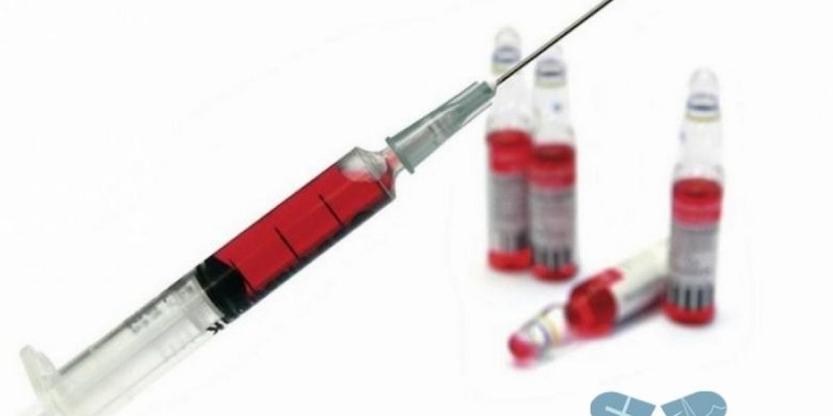
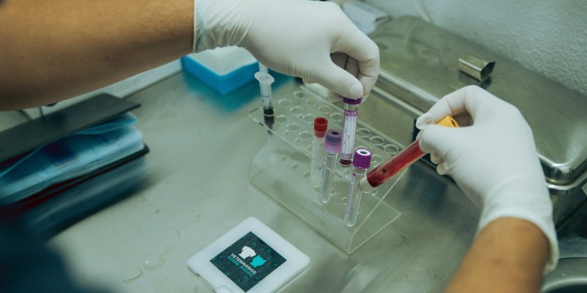
Blood Tests for Dogs That Target Particular Organs
Hyperphosphatemia will increase the manufacturing of parathyroid hormone (PTH) by the parathyroid glands, one of the key steps in growth of renal secondary hyperparathyroidism. Patients of any age may develop CKD, however the best incidence is in geriatric patients. However, congenital renal diseases, including dysplasia and various glomerulopathies, could produce CKD at very early ages. If you think your canine has kidney failure, don’t attempt to self-diagnose and treat the condition at home. Dr. Brittany Kleszynski is a contract veterinary and medical author who specializes in creating significant content that engages readers and speaks directly to the intended audiences.
Monitoring the condition
Affected canines, however, can reside longer and have a greater high quality of life with proper administration that consists of a particular food regimen, medicines, and sustaining fluid and electrolyte stability in the physique. In most cases, cautious consideration of these findings will enable the clinician to discover out whether total kidney perform is considerably compromised. The IRIS guidelines are a vital tool in staging kidney disease in canine. By understanding the totally different phases and the way they are decided, dog house owners can be higher outfitted to make informed choices about their pet’s health.
Kidney Disease in Dogs: Signs, Symptoms, and Treatment
If this isn't practical, then the team might carry out the readings throughout the same visit. In that case, the goal is to provide your canine a significant interval of downtime in between readings. Following initiation or enhance of ACE inhibitor dosage, delicate increases in BUN and creatinine could additionally be noted. Monitor mild will increase that do not trigger uremia; however, reduce or discontinue the dosage if azotemia, accompanied by uremia, considerably increases, which suggests the ACE inhibitor has brought on a big decrease in GFR.
Diagnostics to support patient care
Other symptoms don’t usually turn out to be apparent till about two-thirds of the kidney tissue is destroyed. So, within the case of CKD, the injury may have begun months or even years earlier than the proprietor notices. Because of this, it’s frequent for the indicators of kidney disease in dogs to look like they came out of the blue when in fact, the kidneys have been struggling for a very long time. If your dog has been identified with kidney illness, you need to know their stage. This article will discuss how veterinarians stage kidney illness in dogs, and how to finest perceive the International Renal Interest Society (IRIS) tips.
It decreases due to insulin overdose, insulinoma, islet cell hyperplasia (uncommon), acetonemia/pregnancy toxemia, acute febrile illness, and idiopathically (in certain dog breeds). It decreases in ruminants due to dietary deficiency, both acute (grass staggers) or chronic, and diarrhea (uncommon). Most veterinary laboratories provide a basic panel of checks, which represents a minimal investigation relevant to most basic situations. For https://Tempaste.Com small animals, a typical panel contains total protein, albumin, globulin (calculated as the difference between the first two analytes), urea, creatinine, ALT, and alkaline phosphatase (ALP). In addition, a yellow colour seen in the plasma should be thought of a sign to measure bilirubin. Coagulation exams usually mirror disseminated intravascular coagulation (DIC).18 Anemia is in all probability not present initially, but as merozoites invade RBCs, hemolysis outcomes and would possibly warrant transfusion.
Table 3.10
Resting the pancreas is recommended only if the animal vomits uncontrollably (ie, the animal vomits regularly and violently despite applicable antiemetic therapy). In fact, early nutritional assist is taken into account a key part of successful remedy of human patients with extreme pancreatitis. Animals that vomit ought to be handled with an antiemetic, corresponding to maropitant (NK1 antagonist), ondansetron, or dolasetron (HT3 antagonists), or in most animals a mixture of both. Even animals that do not actively vomit could benefit from such antiemetic assist, as a outcome of they might be nauseated, resulting in poor or absent urge for food. Metoclopramide just isn't thought-about efficient as an antiemetic agent and shouldn't be utilized in these animals. More advanced imaging strategies corresponding to contrast-enhanced ultrasonography, computed tomography, or magnetic resonance imaging usually are not but routinely used for the diagnosis of pancreatitis in canine and cats, though they might maintain promise for the long run.
Tests for Pancreatic Disease:
Site choice depends on the species involved and whether chemical restraint is required. The right jugular vein (Figures 1 and 2) is commonly sampled as it's larger than the left jugular vein in most avian species (Campbell and Ellis, 2007). Relatively large volumes could be drawn, although haematomas can usually form as a outcome of lack of surrounding subcutaneous tissue (Samour, 2006). The jugular may be found underneath a featherless tract of pores and skin known as the apterium, which allows for good visualisation in most species; nonetheless, the jugular apterium is noticeably absent in pigeons and waterfowl (Chitty and Lierz, 2008). Care should be taken regarding the amount of blood that can be safely withdrawn from a patient, particularly in small sick birds (Samour, 2006).
automatic biochemistry analyzerSpotchem™ EZ VET
She completed her ECZM residency in avian medication and surgical procedure at Great Western Exotics in 2022. Ashton gained her certificates of superior veterinary follow in zoological drugs in 2020 and became a complicated practitioner in zoological drugs in 2021. Heteropenia is usually caused by artefacts (Samour et al., 2015); if pathological, it is usually accompanied by a degenerative left shift because of decreased manufacturing or overwhelming demand (Stacy et al., 2022). Heterophilia can result from a stress response or infectious or non-infectious causes of irritation (Chitty, 2018). A left shift and poisonous modifications can be seen in circumstances of heterophilia with more severe inflammatory disease, for example bacterial sepsis (Campbell and Ellis, 2007).
Featured tests, test kits, and single slides
Albumin is decreased as a outcome of decreased manufacturing resulting from liver illness, elevated loss via kidney or gastrointestinal tract disease, and elevated use in continual irritation, for example (Harr, 2006). Elevated alpha and beta globulins usually indicate acute inflammation or an infection, whereas elevated gamma globulins can point out a extra chronic inflammatory or infectious process (Samour et al., 2015). The intensity of color is in part associated to the amount of urine collected and laboratorio analises clinicas veterinaria focus of urine produced; due to this fact, it should be interpreted in context of urine particular gravity. Abnormal urine shade could also be brought on by presence of endogenous or exogenous pigments, nevertheless it does not provide specific data. Interpretation of semiquantitative reagent strips, that are colorimetric exams, requires information of urine shade as a outcome of discolored urine could lead to a false-positive outcome. Urine was collected during surgery by cystocentesis or instantly after surgery (T1) by aseptic catheterization, and at T24 and T48 by voided midstream sample.